Bitewing X-rays
Show the crowns of upper and lower back teeth and the areas between them, helping assess spaces that are difficult to see directly.

General Dentistry
Digital imaging helps your dentist see details that are not visible during a clinical exam—and explain what those details mean for your care.
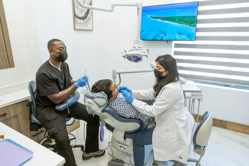
A clearer clinical picture
A visual exam reveals a great deal, but some tooth surfaces, roots, bone, and supporting structures cannot be assessed fully by sight alone. Dental X-rays can provide additional information when your dentist is evaluating symptoms, monitoring oral health, or planning care.
The type and timing of imaging are based on your individual needs. Your dentist considers your health history, current exam, symptoms, prior images, age, and treatment plan before recommending an X-ray.
Imaging options
Your dentist selects the imaging view that best supports the clinical question—not every patient needs every type.
Useful clinical context
Images can show areas between teeth, beneath restorations, around roots, and within supporting bone that are not fully visible.
Diagnostic detail can help your dentist compare care options and plan procedures with more information.
When clinically appropriate, current images can be compared with prior records to monitor areas of interest.
What to expect
We review your symptoms, health history, current exam, and available prior images.
Your dentist recommends the image or series that addresses the specific clinical need.
A sensor or imaging unit is positioned carefully while you remain still for the image.
Your dentist examines the result and explains relevant findings in the context of your oral health.
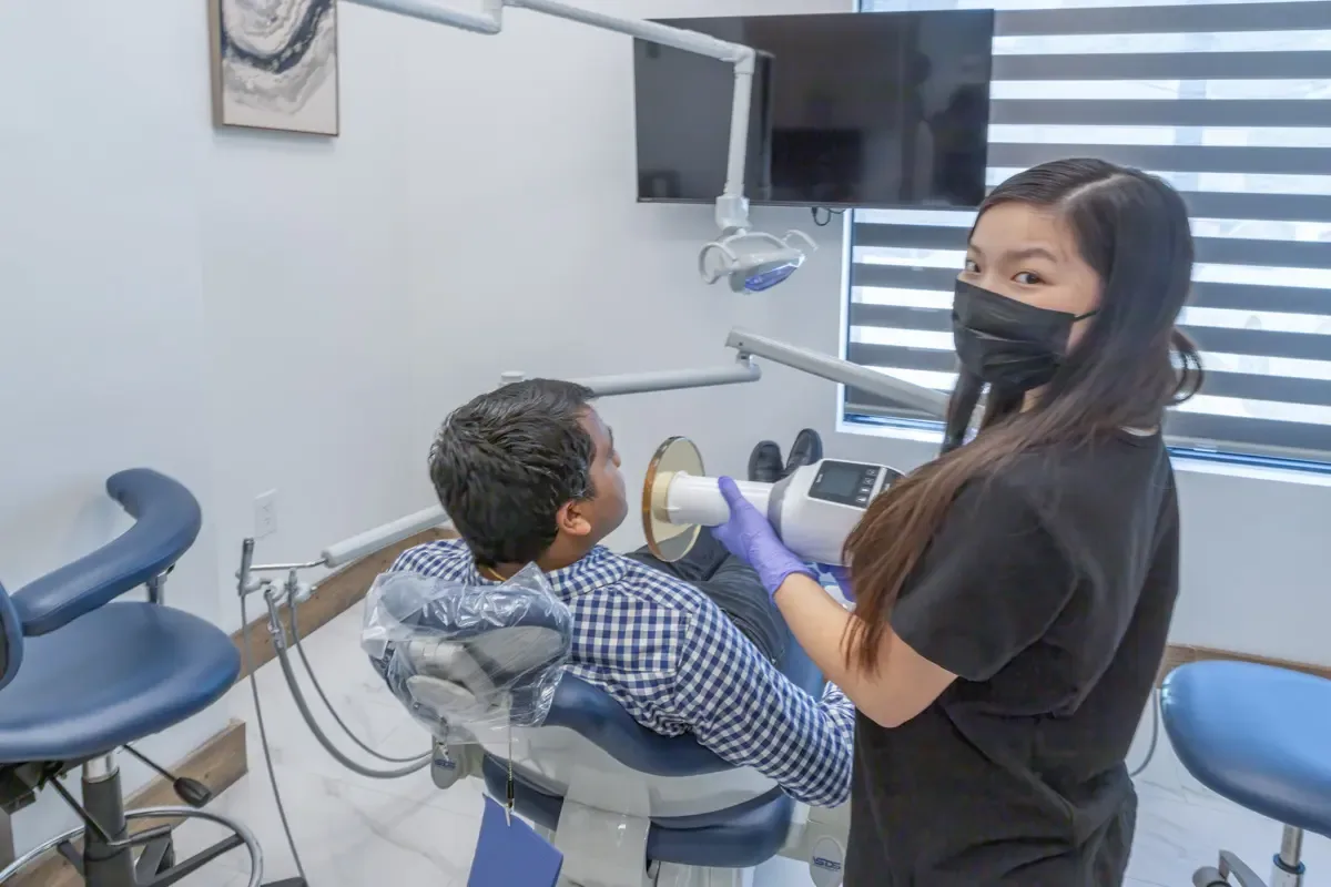
Individualized decisions
Dental imaging is not one-size-fits-all. Recommendations are based on the information your dentist needs for diagnosis, monitoring, or treatment planning, while considering your history and available records.
Need-based recommendations
Image type and timing are matched to your clinical situation.
Digital workflow
Images can be viewed on screen and discussed with you during the visit.
Clear explanations
We connect findings to the exam and the options being considered.
Common questions
Your dentist can answer questions in the context of your age, health history, and current oral health needs.
X-rays may be recommended to evaluate areas that cannot be seen fully during a visual exam, investigate symptoms, monitor a known concern, or support treatment planning.
There is no single schedule for everyone. Timing depends on your age, health and dental history, symptoms, current exam, risk factors, prior images, and treatment needs.
Tell the dental team about relevant medical history and whether you are pregnant or may be pregnant so the dentist can consider that information when deciding what imaging is appropriate.
Some intraoral images require a small sensor or plate to rest inside the mouth. We position it carefully and can adjust when possible if you have a sensitive gag reflex or discomfort.
Digital images can be displayed on screen. Your dentist can point out relevant anatomy and explain how the findings relate to the examination and proposed next steps.
Continue your care
Shape Dental · Georgetown, TX
Book an appointment to discuss symptoms, preventive care, or imaging recommended for your treatment plan.
Most dental plans reset on January 1, and unused benefits usually don’t carry over. If you haven’t had your cleaning and checkup this year, your plan may already cover it.
Not sure what’s left on your plan? Give us a call — we’re happy to help you check.